Bone mineral density of skeletal remains: Discordant results between chemical analysis and DXA method |
| |
| Affiliation: | 1. Department of Medical Laboratory Sciences, University of Delaware, Newark, DE, USA;2. Diabetes and Metabolic Research Center, Christiana Care Health System, Newark, DE, USA;3. Diabetes and Metabolic Diseases Center, Christiana Care Health System, Wilmington, DE, USA;4. Biostatistics Core Facility, University of Delaware, Newark, DE, USA;5. Biomedical Research & Analysis Laboratory, Nemours Children’s Specialty Care & Mayo Clinic College of Medicine, Jacksonville, FL, USA |
| |
| Abstract: | 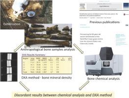
Dual-energy X-ray absorptiometry (DXA) scanning is a gold standard for bone mineral density measurement and diagnosis of primary and secondary osteoporosis in living persons. DXA is becoming widespread when analysing archaeological material, and is considered to provide an accurate diagnosis of osteoporosis in skeletal samples.The aim of this study was to explain the differences in results between bone mineral density (obtained with DXA) and chemical determination of calcium and phosphorus concentrations in skeletal remains. We examined bone mineral density (BMD) and mineral content of femoral bone samples exhumed from mass graves of the Second World War. BMD was determined by Hologic QDR 4500 C (S/N 48034) Bone Densitometer. Concentrations of calcium and phosphorus were determined with AAS (Atomic absorption spectroscopy) and UV/VIS (Ultraviolet–visible) spectroscopy.The results obtained in this study do not support the hypothesis according to which BMD measured by DXA scan has positive correlation with chemically determined concentrations of calcium and phosphorus in bones, especially in acidic soils where there was significant impact of diagenesis observed. |
| |
| Keywords: | Bone density DXA scan Calcium and phosphorus concentration Skeletal remains |
| 本文献已被 ScienceDirect 等数据库收录! |
|