Tracking Multiorgan Treatment Response in Systemic AL-Amyloidosis With Cardiac Magnetic Resonance Derived Extracellular Volume Mapping |
| |
| Affiliation: | 1. National Amyloidosis Centre, University College London, Royal Free Campus, London, United Kingdom;2. Center for Diagnosis and Treatment of Cardiomyopathies, Cardiovascular Department, Azienda Sanitaria Universitaria Giuliano-Isontina (ASUGI), University of Trieste, Italy;3. Cardiology University Department, IRCCS Policlinico San Donato, Milan, Italy;4. St Bartholomew''s Hospital, London, United Kingdom;5. National Heart, Lung and Blood Institute, National Institutes of Health, Bethesda, Maryland, USA |
| |
| Abstract: | 
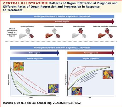
BackgroundSystemic light chain amyloidosis is a multisystem disorder that commonly involves the heart, liver, and spleen. Cardiac magnetic resonance with extracellular volume (ECV) mapping provides a surrogate measure of the myocardial, liver, and spleen amyloid burden.ObjectivesThe purpose of this study was to assess multiorgan response to treatment using ECV mapping, and assess the association between multiorgan treatment response and prognosis.MethodsThe authors identified 351 patients who underwent baseline serum amyloid-P-component (SAP) scintigraphy and cardiac magnetic resonance at diagnosis, of which 171 had follow-up imaging.ResultsAt diagnosis, ECV mapping demonstrated that 304 (87%) had cardiac involvement, 114 (33%) significant hepatic involvement, and 147 (42%) significant splenic involvement. Baseline myocardial and liver ECV independently predict mortality (myocardial HR: 1.03 [95% CI: 1.01-1.06]; P = 0.009; liver HR: 1.03; [95% CI: 1.01-1.05]; P = 0.001). Liver and spleen ECV correlated with amyloid load assessed by SAP scintigraphy (R = 0.751; P < 0.001; R = 0.765; P < 0.001, respectively). Serial measurements demonstrated ECV correctly identified changes in liver and spleen amyloid load derived from SAP scintigraphy in 85% and 82% of cases, respectively. At 6 months, more patients with a good hematologic response had liver (30%) and spleen (36%) ECV regression than myocardial regression (5%). By 12 months, more patients with a good response demonstrated myocardial regression (heart 32%, liver 30%, spleen 36%). Myocardial regression was associated with reduced median N-terminal pro-brain natriuretic peptide (P < 0.001), and liver regression with reduced median alkaline phosphatase (P = 0.001). Changes in myocardial and liver ECV, 6 months after initiating chemotherapy, independently predict mortality (myocardial HR: 1.11 [95% CI: 1.02-1.20]; P = 0.011; liver HR: 1.07 [95% CI: 1.01-1.13]; P = 0.014).ConclusionsMultiorgan ECV quantification accurately tracks treatment response and demonstrates different rates of organ regression, with the liver and spleen regressing more rapidly than the heart. Baseline myocardial and liver ECV and changes at 6 months independently predict mortality, even after adjusting for traditional predictors of prognosis. |
| |
| Keywords: | cardiac magnetic resonance extracellular volume mapping systemic AL amyloidosis AL" },{" #name" :" keyword" ," $" :{" id" :" kwrd0030" }," $$" :[{" #name" :" text" ," _" :" light chain CMR" },{" #name" :" keyword" ," $" :{" id" :" kwrd0040" }," $$" :[{" #name" :" text" ," _" :" cardiac magnetic resonance ECV" },{" #name" :" keyword" ," $" :{" id" :" kwrd0050" }," $$" :[{" #name" :" text" ," _" :" extracellular volume FLC" },{" #name" :" keyword" ," $" :{" id" :" kwrd0060" }," $$" :[{" #name" :" text" ," _" :" free light chain SAP" },{" #name" :" keyword" ," $" :{" id" :" kwrd0070" }," $$" :[{" #name" :" text" ," _" :" serum amyloid P component |
| 本文献已被 ScienceDirect 等数据库收录! |
|

