Robust whole-brain segmentation: Application to traumatic brain injury |
| |
| Affiliation: | 1. Biomedical Image Analysis Group, Department of Computing, Imperial College London, UK;2. The Neurodis Foundation, CERMEP, Lyon, France;3. Division of Brain Sciences, Faculty of Medicine, Imperial College London, UK;4. The PET Centre, Division of Imaging Sciences and Biomedical Engineering Kings College London, St Thomas Hospital, London, UK;5. MedTech West, Institute of Neuroscience and Physiology, University of Gothenburg, Sweden;6. Knowledge Intensive Services, VTT Technical Research Centre of Finland, Tampere, Finland;7. University Division of Anaesthesia, University of Cambridge, Addenbrooke’s Hospital, Cambridge, UK |
| |
| Abstract: | 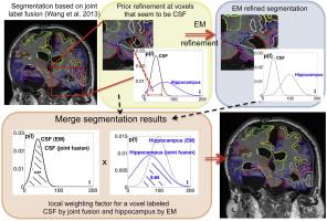
We propose a framework for the robust and fully-automatic segmentation of magnetic resonance (MR) brain images called “Multi-Atlas Label Propagation with Expectation–Maximisation based refinement” (MALP-EM). The presented approach is based on a robust registration approach (MAPER), highly performant label fusion (joint label fusion) and intensity-based label refinement using EM. We further adapt this framework to be applicable for the segmentation of brain images with gross changes in anatomy. We propose to account for consistent registration errors by relaxing anatomical priors obtained by multi-atlas propagation and a weighting scheme to locally combine anatomical atlas priors and intensity-refined posterior probabilities. The method is evaluated on a benchmark dataset used in a recent MICCAI segmentation challenge. In this context we show that MALP-EM is competitive for the segmentation of MR brain scans of healthy adults when compared to state-of-the-art automatic labelling techniques. To demonstrate the versatility of the proposed approach, we employed MALP-EM to segment 125 MR brain images into 134 regions from subjects who had sustained traumatic brain injury (TBI). We employ a protocol to assess segmentation quality if no manual reference labels are available. Based on this protocol, three independent, blinded raters confirmed on 13 MR brain scans with pathology that MALP-EM is superior to established label fusion techniques. We visually confirm the robustness of our segmentation approach on the full cohort and investigate the potential of derived symmetry-based imaging biomarkers that correlate with and predict clinically relevant variables in TBI such as the Marshall Classification (MC) or Glasgow Outcome Score (GOS). Specifically, we show that we are able to stratify TBI patients with favourable outcomes from non-favourable outcomes with 64.7% accuracy using acute-phase MR images and 66.8% accuracy using follow-up MR images. Furthermore, we are able to differentiate subjects with the presence of a mass lesion or midline shift from those with diffuse brain injury with 76.0% accuracy. The thalamus, putamen, pallidum and hippocampus are particularly affected. Their involvement predicts TBI disease progression. |
| |
| Keywords: | Traumatic brain injury Magnetic resonance imaging Multi-atlas segmentation Brain image segmentation Expectation–maximisation |
| 本文献已被 ScienceDirect 等数据库收录! |
|